
A 50-year-old man is referred by his GP for the evaluation of headache and very high blood pressure. In the past, he was investigated for obstructive uropathy by Urologist but he was lost to follow up.
He had no history of hypertension and was not on any regular medications.
Physical examination findings are as follows:
Pulse rate – 82/min
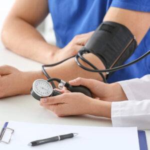
Blood pressure – 232/128 mm Hg
Heart sounds – no murmurs
Lungs – no adventitious sounds. Bilateral equal air entry
Abdominal examination – soft, non tender. No organomegaly.
CNS: GCS 15/15, no focal neurology, no papilloedema on fundoscopy
Secondary hypertension should be suspected in patients who exhibit certain clinical characteristics or have specific risk factors.
- Early Onset or Severe Hypertension: If hypertension is diagnosed at a young age (before 40 years) or if blood pressure levels are unusually high, secondary hypertension should be considered.
- Hypertension Resistant to Treatment: If a patient’s blood pressure remains uncontrolled despite the use of multiple antihypertensive medications, secondary hypertension should be suspected.
- Abrupt Onset of Hypertension: Hypertension that develops suddenly without any apparent reason might be indicative of secondary hypertension.
- Family History of Early-Onset Hypertension: A family history of hypertension, especially if it presents at a young age, may suggest a possible genetic cause or a familial form of secondary hypertension.
- Presence of Associated Symptoms: Certain symptoms may be indicative of specific causes of secondary hypertension. For example:
- Severe headache, palpitations, and sweating may suggest pheochromocytoma.
- Muscle weakness, fatigue, and polyuria may indicate primary aldosteronism.
- Weight gain, edema, and reduced urine output could be related to kidney disease.
- New-Onset Hypertension in Older Individuals: In older patients, the development of hypertension for the first time should raise suspicion of secondary causes.
- Abnormal Laboratory Findings: Any abnormal results from routine blood tests, such as electrolyte imbalances, abnormal kidney function, or abnormal urine analysis, can point to potential secondary hypertension causes.
- Known Underlying Medical Conditions: Certain medical conditions are associated with an increased risk of secondary hypertension. These include chronic kidney disease, endocrine disorders (e.g., diabetes, Cushing’s syndrome, hyperthyroidism), and sleep apnea.
- Medication-Induced Hypertension: Some medications, such as oral contraceptives, nonsteroidal anti-inflammatory drugs (NSAIDs), and certain antidepressants, can cause or worsen hypertension.
- Hypertension in Pregnancy: Hypertension that develops during pregnancy or worsens during pregnancy (gestational hypertension or preeclampsia) may indicate an underlying secondary cause.
- Flash pulmonary edema or widespread atherosclerosis: renal artery stenosis
- Cold legs: poor distal perfusion due to aortic coarctation
- Swelling and hypertension in a pregnant patient: pre-eclampsia
- Oedema and foamy urine in a non-pregnant patient: nephrotic syndrome
- History of renal impairment, prostatic enlargement, previous urethral instrumentation, or renal calculi: obstructive uropathy or chronic kidney disease
- Family history of polycystic kidney disease, intracranial aneurysms, or subarachnoid haemorrhage in a young patient with hypertension (age 20-34 years): polycystic kidney disease
- Phaeochromocytoma: episodic symptoms consistent with a hyper-adrenergic state, such as panic attacks, sweating, palpitations, and abdominal cramps
- Symptoms of low potassium: hyperaldosteronism (although most patients are normokalemic)
- Typical symptoms of Cushing’s syndrome: depression, weight gain, hirsutism, easy bruising, and low libido
- Excess thyroxine: heat intolerance, sweating, palpitations, and weight loss; low thyroxine levels: lethargy, constipation, weight gain, and depression
- Symptoms of bone pain, paraesthesia, and myalgia: hyperparathyroidism
- Excessive daytime sleepiness in an obese patient, along with erectile dysfunction and restless sleep: obstructive sleep apnoea
- Toxic causes: use of oral contraceptive pill, non-steroidal anti-inflammatory drugs, or chronic alcohol excess.
Physical findings suggestive of a secondary cause include:
- Renal bruits: renal artery stenosis
- Enlarged kidneys, hepatomegaly, or hernia: polycystic kidney disease
- Arteriovenous fistulae: end-stage kidney disease
- Flank tenderness or prostatic enlargement on rectal examination: obstructive uropathy
- Facial or limb oedema in a pregnant patient: pre-eclampsia; in a non-pregnant patient: nephrotic syndrome
- Radio-femoral delay, disparity in blood pressure readings between arms, systolic or continuous cardiac murmurs, weak or impalpable distal pulses: coarctation of the aorta
- Features of Cushing’s syndrome: moon face, thin arms and legs, truncal obesity, striae, and skin thinning
- Hypothyroidism: isolated eyelid oedema, dry skin, thick tongue; Hyperthyroidism (Graves’ disease): exophthalmos, proptosis, and lid lag
- Deposition of calcium inside the iris or palpable jaw tumors: hyperparathyroidism
- Obesity: obesity hypoventilation syndrome; obesity, maxillomandibular abnormalities, and macroglossia: obstructive sleep apnoea
Initial Investigations:
Electrocardiogram (ECG): To look for signs of previous myocardial infarction or left ventricular hypertrophy, which is a key prognostic factor.
Chest X-ray: To check for cardiomegaly, widening of the left subclavian border, and a double bulge at the site of the aortic knuckle, which may be seen in coarctation of the aorta, along with notching of the ribs due to large collateral circulation.
Blood Tests:
- Urea, electrolytes, and creatinine: To assess kidney function.
- Random blood sugar and serum cholesterol: As part of overall cardiovascular risk assessment.
- Fasting blood sugar (if diabetes is suspected): To diagnose diabetes.
- Potassium levels: To investigate hyperaldosteronism, although they are usually normal.
Urine Dip Test: To check for glycosuria and proteinuria, and the presence of casts to determine underlying renal causes such as glomerulonephritis or nephrotic syndrome.
Subsequent Investigations (if clinical suspicion of secondary hypertension is high):
Blood Tests:
- Plasma renin and aldosterone levels (if hyperaldosteronism is suspected).
- Adrenal vein sampling (to compare the ratio of renin to aldosterone in each kidney) if hyperaldosteronism is suspected.
- Plasma renin activity (elevated in most patients with renal artery stenosis) – a good screening test.
- Liver function tests (if chronic alcohol excess and liver dysfunction are suspected).
- Thyroid function tests (if hyper- or hypothyroidism is suspected).
- Serum calcium levels (if hyperparathyroidism is a possibility).
Urine Tests:
- 24-hour urine collection: For measuring catecholamines (to exclude a phaeochromocytoma) and protein levels (in suspected pre-eclampsia or nephrotic syndrome). Spot urine creatinine ratio can offer comparable results for pre-eclampsia and nephritic syndrome.
Imaging:
- Ultrasound of kidneys and adrenal glands: To visualize kidney size and structure, as well as adrenal abnormalities.
- CT of adrenals: To localize a phaeochromocytoma if urinary catecholamines are suggestive of the diagnosis.
- MRI: To investigate renal artery stenosis if a renal angiogram is contraindicated, and for imaging adrenal glands to localize tumours in hyperaldosteronism or phaeochromocytoma.
Biopsy:
- Renal biopsy: A definitive test to determine the underlying cause of nephrotic syndrome.
These investigations help in identifying the specific cause of secondary hypertension and guide the appropriate treatment plan.